Buscador de Empresas y Servicios lasguias.com
Portales para mejorar el SEO Local y la venta online
Directorio Incluir empresa Blog de LasGuias Contactar Web corporativa
PODOLOGOS EN BARCELONAPODOLOGOS ▷ BARCELONA ▷ BARCELONAListado de empresas
|
Actividades relacionadas en la temática
Centros de salud: · Salud mental y psiquiatrica: centros sanitarios en Barcelona · Centros asistenciales de dia en Barcelona · Centros de salud en Barcelona · Centros medicos en Barcelona · Residencias de reposo en Barcelona · Sanatorios en Barcelona · Buscar podologos en RinconPymes. |
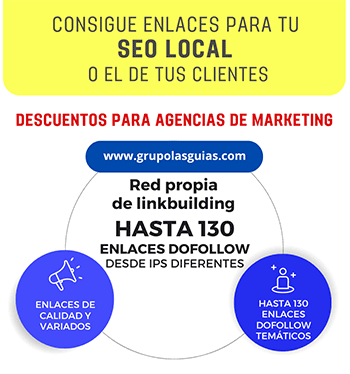
🔗 ENLAZAMOS A TU WEB PARA MEJORAR TU SEO EN GOOGLE 📈
El Buscador de Las Guías pertenece a Grupo Lasguías, especialistas en el posicionamiento web y consta de un grupo de directorios con enlaces dofollow. Saber más sobre cómo conseguir enlaces válidos para el posicionamiento en Google. Nuestro Directorio de empresas lasguias.com está especializado en empresas españolas y en mejorar el posicionamiento de páginas web en Google y otros buscadores. Además, Grupolasguias ofrece otros servicios, como la traducción de páginas web, documentos y productos de venta online,así como la creación de posts o artículos de negocios en nuestro blog, servicio conocido también como escritor de contenidos web. En el caso de querer comprar enlaces en blogs pulse en el link anterior para saber los blogs que administramos y sus temáticas.
Copyright (c) lasguias.com .- Todos los derechos reservados. Publicidad, Creación y Desarrollo, y Servicios de Búsqueda en Internet.
[ Quienes Somos ]
[ Entrar a mi correo @lasguias.com ]
[ CONTACTO ]
[ Mapa de la web ] - AVISO LEGAL - Política de Privacidad -
Política de Cookies